Why gaps appear between teeth and implants
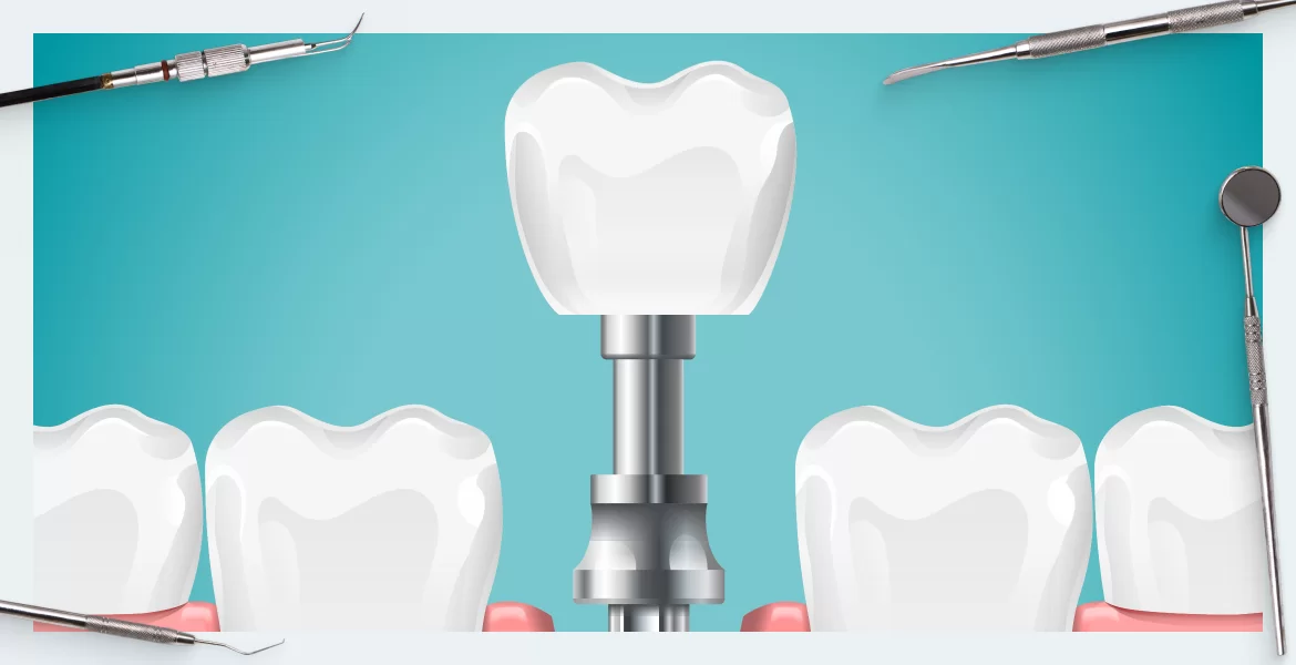
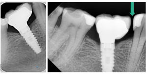
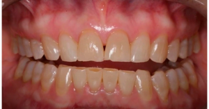
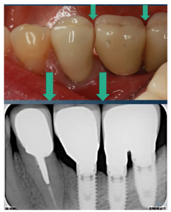
Today we will deal not only with the cause of the gaps between the living tooth and the crown (they are open proximal contacts), but we will also analyze recommendations for their elimination. This quite in general practice in a dead end. Fortunately, it rarely causes critical problems, but food debris can accumulate in the gap injuring the gum and the paratond of the neighboring tooth, as well as promoting the development of tooth decay. The main problem for many experts is the surprise of this phenomenon. After healing, everything looks great and correct as in the top picture below, but after a few years there is a gap, see the example in the lower photo below.
The gap between tooth and implant restoration – when and how often it happens
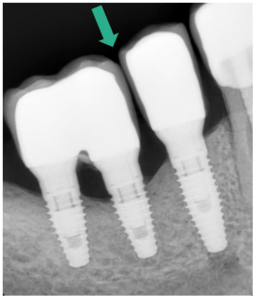
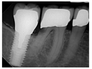
Let’s analyze the clinical case from the above photos. When the implant was installed in 2013, the X-ray looked like this, see the left photo below. The patient was satisfied, but almost did not come to check-ups. Four years later, there was a gap between the 5th and 6th teeth. The sixth tooth is a restoration on the implant, and the fifth living tooth with a restoration from a composite seal.
At the time of installation, both the photoprotocol and the X-rays were taken. Flosses were also checked, and everything was within the normal range. However, the result is clear. We will further analyze the causes of gaps; here we note that this phenomenon is quite frequent. In most cases, it occurs in single implants and in fairly young patients.
Unfortunately, this phenomenon is not well understood and described, as it rarely causes great discomfort in patients. However, there are reviews and studies on this.
This publication brings together five studies where patient observations ranged from 2.2 to 12 years. The studies studied here were:
- Etiology (origin);
- Causes;
- Frequency of occurrence;
- Correction – open contacts between implants and live teeth.
The number of observations and studies here does not qualify it as a large-scale study, but the general patterns and methods of problem elimination are made qualitatively.
This is what the statistics show: Problems begin to appear at the time of 3 months and open contact proximal surfaces appear at a frequency of 34% to 66%. The degree of expression is different and there is a difference between the upper and lower jaws. On the lower jaw, the cracks appear much more often, but the cause is not fully understood.
It was also noted that gaps between the teeth in patients are more frequent and more pronounced:
- with bruxism and other hyperfunctions of the jaw system;
- With reflux, when acid from the stomach enters the mouth cavity, the acidic environment accelerates the enamel removal, which accelerates the formation of the gap.
What are the causes of gaps between teeth and implants
There are only three reasons for the appearance of a gap between the implant and the tooth:
- Mesic drift.
- A zuboalveolar extension.
- Elasticity of the jaw.
The main reason on which we will dwell is the mesic drift. This is the most studied and proven factor, the rest affect only to a small extent.
What is mesic drift?
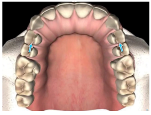
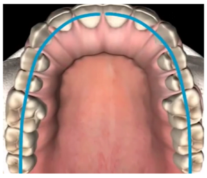
The phenomenon of mesial drift is well known to orthodontists and, to a lesser extent, general dentists. The essence of the phenomenon is that all the upper and lower jaw teeth are slightly tilted away from the distal part of the skull (from the back of the head toward the nose). That is, they have a mesial tilt – this tilt is expressed differently, but is always present.
In addition to the initial inclination over the course of life, the row of teeth is gradually moving forward. Moreover, in the process of chewing, and also in the ordinary life, we very often squeeze our teeth. Ligaments and muscles create a force vector that further increases the mesic slope.
The working and lateral surfaces of the teeth are erased during the life process. Mesic drift in some ways compensates for this phenomenon. The teeth are pushed forward and pressed against each other as they rub. While a rigidly attached jaw titanium implant does not participate in drift.
Over time, the adjacent tooth, located behind it along the drift vector, “floats away” and a gap is formed.
There are forces that act on the dentition distally, but these forces are five times less than the medial ones. That is why distal drift is uncommon, at least not properly described in the world practice.
Whereas mesial drift is studied by orthodontists and other specialists.
In terms of eliminating the gap between the living tooth and the implant, the multi-unit dental implant platform is much more convenient. The bridge with crowns can be removed quite easily, and composite material can be added to fill the gap and put the bridge back on. We’ll go into more detail about the removal technique later. There is a problem though, the multi-unit system is almost never used for single restorations.
Tooth abrasion – evolutionary norm or anomaly?
The process of natural tooth abrasion is also important in the study of the phenomenon of mesial drift. Living teeth have a micromobility and during chewing can shift, deviate and diverge from each other by up to 100 microns in the vestibulo-oral direction.
The tooth also moves vertically and can “sink” into the jawbone by up to 25 microns when the jaws are compressed. All this micromobility leads to the erasure of the occlusal surfaces of the teeth, but also the lateral (proximal) surfaces in the contact areas with neighboring teeth.
It is also known that the nature of the contact surface of adjacent teeth changes with age. In young teeth it is cusp-like, and in older teeth it is flat.
As we already know, there are no gaps between the teeth, and patients do not complain about food getting stuck between the teeth. Unless the tooth is affected by decay.
In fact, with age, teeth rub and fit together. In an ideal case, the teeth resource and compensatory mechanism should be enough for the rest of life.
Unfortunately, most people lose some living teeth much earlier.
The issue of mesial drift and tooth erasability has been studied by many, such as Dr. Gottlieb and other specialists.
We are interested to find out how unique this phenomenon is. We will start the analysis with an interesting study of our fellow biologists from 2012.
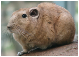
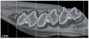
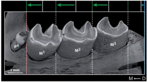
They conducted research on the crested rat. The animal was not chosen by chance – it is one of the oldest mammals. And there are sufficient fossil remains of teeth and jaws of these animals in the storerooms of Belgium and other countries. So, studying modern rats together with paleontological data it is possible to make a good sampling and to trace the phenomenon of abrasion and mesial drift of teeth.
Tomographic, radiographic, and photographic data were used for the work. This made it possible to understand, in principle, how this process takes place.
Rattlesnake rats are rodents, and it is logical that their teeth are designed to chew hard food, plus they are used as a tool for building a home. It is logical that they wear down and grind very hard. The phenomenon of mesial drift is significantly stronger in these animals.
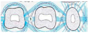
It was interesting to find out exactly how and at what expense the mesial drift takes place. It transpired that the ligamentous apparatus is actively involved in the process. As we have already mentioned in the series of articles on soft tissue integration, a living tooth is surrounded by the gingival cuff. Underneath the epithelium layer is a complex of multidirectional connective tissue fibers (collagen fibers).
In this case, we are interested in the group of transseptal fibers (marked with number 1 in the illustration below), which connect one well to another and all the teeth appear to be connected in something remotely resembling a chain.
The mechanical stress of chewing pushes the teeth forward. The entire ligament apparatus helps to distribute the load on the entire tooth row almost synchronously and presses them against each other. However, if the teeth just leaned and pressed against each other and then returned to their original position, sooner or later the safety margin would run out. The lateral surfaces would wear away and gaps would inevitably appear. However, this does not happen, thanks to concomitant processes in the alveolar bone.
In the mesial plane (in front of the tooth) the bone is resorbed and the tooth roots occupy the vacated space. At the same time in the distal part, the free space is filled with new bone tissue. So the tooth is fixed in its new position. The teeth literally drift and maintain a plus-minus stable position relative to each other.
This is why there are no gaps between the teeth, even as the proximal (lateral) surfaces erode. Obviously, in the case of an implant, its neighbor on the front side “floats away,” and if there is a tooth on the distal side, its drift is stopped by the implant itself.
This process is smooth and takes place over an extended period of time. The rate of mesial drift is affected by the load on the teeth as well as by natural abrasion.
Now let’s look at the process of abrasion and mesial drift in humans. To do this, let’s turn to the research of Japanese specialists. This is a joint work of anthropologists and dentists.
The research team studied the skulls and jaws of people who lived in Japan many thousands of years. They studied the fossils and jaw remains of people who lived 12,000 years ago and 3,000 years ago.
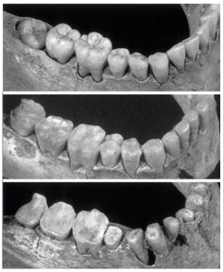
The picture below shows the jaws of Upper Paleolithic people who were hunter gatherers. This is a rather primitive level of development. These people were characterized by eating rather coarse food. Plus, they often softened the skin with their teeth and chewed fibrous parts of plants to obtain bundles of fibers for making tools.
Therefore, the image shows significant abrasion of the occlusal surfaces and pronounced abrasion of the proximal surfaces. From the condition of the teeth you can precisely determine the age of the person.
The picture shows three jaws of people of different ages. The upper picture is the jaw of a very young person, under 20 years of age. The second picture is of a middle-aged man, and the lower picture is the jaw of someone 40-45 years old, an elderly person by Paleolithic standards.
The study of a large number of specimens enabled us to conclude that there is always mesial drift; only the degree of its expression differs. The length of the occlusal arch decreases with age, the teeth are erased, and the dentition is compacted.
The mesial drift is less pronounced in modern people compared to the Upper Paleolithic people. This is not surprising because the load on the maxillary apparatus in the modern world is several times less.
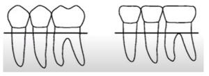
Also during this study, it was found that teeth drift not only in the mesial plane, but also in the coronal plane, see the illustration below. This is also referred to as dental-alveolar motion.
The teeth move upward from the alveolar bone as the occlusal surface is worn away. This process compensates for possible bite problems.
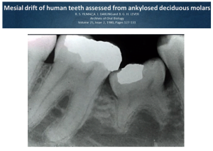
Also, coronal drift violation is well illustrated by the following work. The picture below shows a case of ankylosis of a primary molar.
Ankylosis in this case is a pathological change in the periodontal ligament and the fusion of the tooth roots with the bone. Such a tooth has something in common with a root implant; the restored tooth is excluded from the mesial and coronal drift processes.
The study of such cases also helps to understand the phenomena of mesial and coronal drift. In this case, the ankylosed tooth is a reference point and allows clear tracing of tooth displacement in both planes.
Both bilateral and unilateral cases of ankylosis have been studied. It was found out that healthy teeth participate in the mesial drift, while the ossified molar is an obstacle. The teeth behind it don’t just stop; they get tilted as seen in the picture above.
A full restoration on a root implant behaves similarly to an ankylosed tooth, but its size prevents it from tilting.
The implant does not participate in either the dental-alveolar movement or the mesial drift. Therefore, if the implants were placed at a very young age, in a couple of decades the adjacent teeth will be noticeably higher because they will be advancing and the crown on the root implant is not, not to mention the gaps between the teeth and implants.
Occurrence of open contacts between implants
It would seem that such a phenomenon should not be, but look at this picture. Restoration of the dentition was carried out in 2007, and a patient came in complaining of food getting stuck in 2016. The picture clearly shows the gap between the restored pair and a single implant.
Interestingly, from 2007 to 2015 everything was fine, there was no gap. It appeared literally in six months in 2016. This is not an isolated case, see photo and radiograph below.
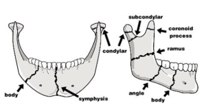
The good news is that these are quite rare cases; the bad news is that the cause of this phenomenon is still unclear. There is speculation that it may be related to the elasticity of the lower jaw.
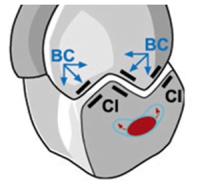
There are areas where the bone is less dense as in the illustration below. These are the lines where the jaws are most likely to break in trauma. There is a possibility that the displacement of implants relative to each other is due to the change in the geometry of the jaw itself during the course of life.
Recommendations for managing open proximal contacts next to implant-based restorations
Although the appearance of the gap itself rarely causes patients to complain, it is not a harmless phenomenon. In the picture below, you can see that food debris has built up in the gap. This led to tooth root decay of the adjacent sixth tooth.
All this will lead to crown replacement, root pulping, etc. The volume of procedures is significant and expensive.
To summarize, we have formulated the following list of recommendations for dentists who manage and regularly examine their patients.
- Inform the patient about the rather high likelihood of a gap and the importance of coming in for preventive check-ups.
- It is recommended that restorations that are relatively easy to repair are created. For example, create splinted structures based on the multi-unit platform. In this system, the restoration is not cemented to the abutment but rather screwed together. It is much easier to remove the restoration and apply an additional layer of composite to compensate for the gap. This involves sandblasting the ceramic surface, applying hydrofluoric acid, that is, making an adhesive preparation, and gluing the composite in place. It is also always better to do splinted restorations rather than several single restorations in a row.
- Before the impression procedure, modify both adjacent contacts with minor recontouring so that they are even in profile and rounded. Grind away undercuts and any roughness.
- If an open contact is found, but there is no ingress or entrapment of food, no treatment is necessary. Observation is sufficient.
- If there is a food jam, the first step is to modify the implant to fill the gap. An augmentation of the adjacent tooth is the second option.
- If there is an open contact with stuck food and decay on the tooth, you need to treat the adjacent tooth with a filling or crown.
- The risk of open contact is higher in patients with bruxism and other disorders that lead to increased tooth abrasion. In this case, the fabrication and wearing of an overnight mouth guard is mandatory!
We hope you have found this article useful. Until future publications …


