Revolutionizing Oral Cancer Detection: AI and Nanotechnology Unite
Introduction: A New Frontier in Oral Cancer Diagnostics
Oral cancer remains a formidable global health challenge, with approximately 390,000 new cases and over 188,000 deaths reported worldwide in 2022 alone. Early detection is paramount, yet traditional diagnostic methods often fall short in identifying the disease at its nascent stages. In a groundbreaking study, researchers from the University of Otago have unveiled a pioneering approach that combines atomic force microscopy (AFM) with artificial intelligence (AI) to detect oral cancer earlier and with greater accuracy. This innovative method promises to transform cancer diagnostics, offering hope for improved patient outcomes and advancing the field of precision medicine.
Key Findings: Harnessing Nanotechnology and AI for Enhanced Detection
The study, published in ACS Nano, highlights the synergistic power of AFM and AI in identifying nanoscale changes on the surface of cancer cells—alterations that traditional diagnostic tools may overlook. By leveraging the high-resolution capabilities of AFM and the pattern-recognition prowess of AI, the researchers achieved a level of diagnostic precision previously unattainable.
- Nanoscale Detection: AFM enables the visualization of minute structural changes on the cell surface, providing a detailed topographical map that reveals early signs of malignancy.
- AI Integration: Advanced AI algorithms analyze the AFM-generated data, discerning subtle patterns and anomalies indicative of cancerous transformations.
- Clinical Implications: This combined approach enhances diagnostic accuracy, potentially allowing for earlier intervention and more effective treatment strategies.
Mechanisms & Technical Pathways: The Science Behind the Innovation
Atomic Force Microscopy (AFM)
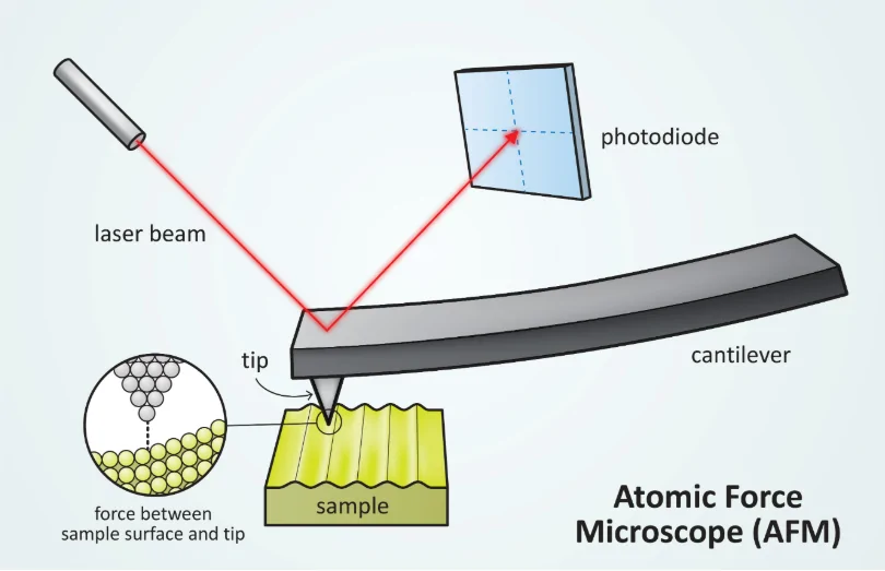
AFM operates by scanning a sharp probe over the cell surface, detecting forces between the probe and the sample to generate high-resolution images.

Illustration of an Atomic Force Microscope (AFM) probe tip. AFMs are so sensitive that they can image individual atoms. /NISE
This technique captures the topography of cells at the nanoscale, revealing structural nuances that may signal the onset of cancer.
Artificial Intelligence (AI) Analysis
AI algorithms process the complex data obtained from AFM, identifying patterns and deviations associated with cancerous cells. Machine learning models are trained on vast datasets to recognize the subtle differences between healthy and malignant cells, enhancing diagnostic precision.
Integration of AFM and AI
The fusion of AFM and AI creates a powerful diagnostic tool that combines detailed imaging with intelligent analysis. This integration allows for the detection of early-stage cancerous changes, facilitating prompt and targeted treatment interventions.
Implications for Professionals: Transforming Clinical Practice
The adoption of this AFM-AI diagnostic method holds significant promise for healthcare professionals:
- Early Detection: Enables clinicians to identify oral cancer at its earliest stages, improving prognosis and survival rates.
- Precision Medicine: Supports the development of personalized treatment plans based on detailed cellular analysis.
- Broader Applications: Potential to extend this diagnostic approach to other cancer types, enhancing early detection across oncology.
- Clinical Integration: Encourages the incorporation of advanced diagnostic technologies into routine clinical practice, elevating the standard of care.
Study Overview: Methodology and Approach
The research team conducted a comprehensive study involving the following steps:
- Sample Collection: Obtained cell samples from patients diagnosed with oral cancer.
- AFM Imaging: Utilized AFM to capture high-resolution images of the cell surfaces, focusing on nanoscale structural features.
- AI Analysis: Applied machine learning algorithms to analyze the AFM data, identifying patterns indicative of malignancy.
- Validation: Cross-referenced AI findings with established diagnostic criteria to assess accuracy and reliability.
This methodological approach underscores the potential of combining nanotechnology and AI to enhance cancer diagnostics.
Data Visualization: Diagnostic Workflow
Conclusion: A Paradigm Shift in Cancer Diagnostics
The integration of atomic force microscopy and artificial intelligence marks a significant advancement in the early detection of oral cancer. By enabling the identification of nanoscale cellular changes, this innovative approach offers a more accurate and timely diagnosis, paving the way for improved patient outcomes. As the medical community continues to embrace technological innovations, such interdisciplinary collaborations will be instrumental in transforming cancer care and enhancing the efficacy of diagnostic methodologies.
Sources
- ScienceDaily – Pioneering method detects oral cancer earlier – April 14, 2025
- ACSPublications – Atomic Force Microscopy for Revealing Oncological Nanomechanobiology and Thermodynamics – March 14, 2025


