Advancements in Understanding and Treating Periodontitis Through Improved Mouse Models
Introduction to Periodontitis and Its Global Impact
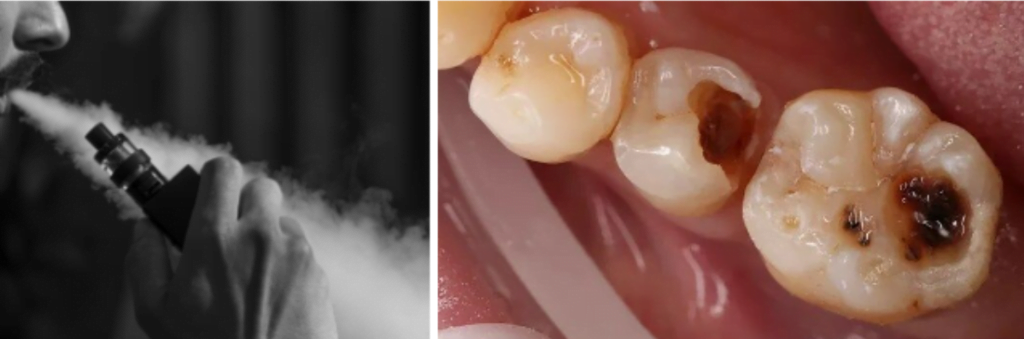
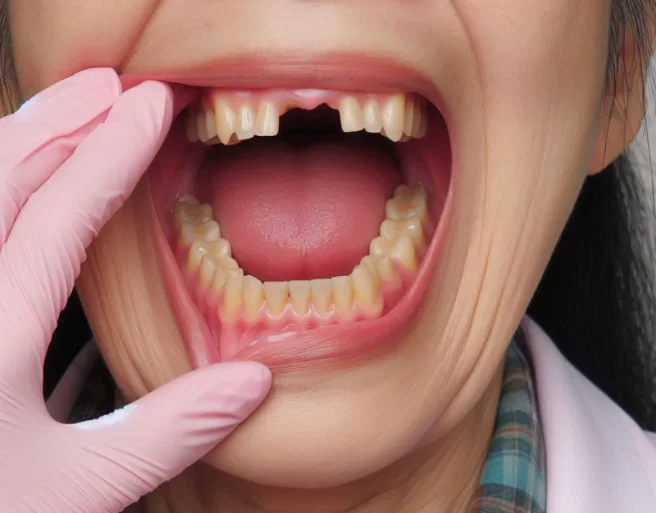
Periodontitis, a severe form of periodontal disease, is a primary cause of tooth loss affecting approximately 20% of adults globally. This inflammatory condition, triggered by bacterial infection of the periodontal tissues, leads to the destruction of the gums, periodontal ligament, and alveolar bone. The prevalence of periodontitis increases with age, making it a significant public health concern as populations worldwide continue to age.

Challenges in Studying Periodontitis
Studying periodontitis directly in humans presents significant challenges due to the complexity of the disease and ethical considerations. Consequently, researchers often rely on animal models to conduct preclinical research. Traditional mouse models, such as the ligature-induced periodontitis model, have been instrumental in understanding the disease. However, these models have limitations in capturing the complete pathology of periodontitis due to restricted tissue sampling.
Innovations in Mouse Models for Periodontitis Research
Researchers from Tokyo Medical and Dental University (TMDU) have recently developed an improved mouse model that addresses the limitations of previous models. This innovative approach utilizes a triple ligature method on the upper left molar of male mice, enhancing the analysis of multiple periodontal tissue types. The improved model significantly increases the yield of peri-root tissue, allowing for a more comprehensive examination of the cellular and molecular mechanisms of periodontitis.
Detailed Analysis of Periodontal Tissue Components
The enhanced mouse model facilitates the simultaneous analysis of various periodontal tissue components, including gingiva, periodontal ligament, alveolar bone, and cementum. This comprehensive approach enables a deeper understanding of the disease’s progression and the identification of key molecular pathways involved in inflammation and bone destruction.
Key Findings on Inflammatory Pathways
Utilizing the improved model, the TMDU research team investigated gene expression changes in periodontal tissues over time, focusing on inflammation-related genes. They discovered a significant increase in the expression of the Il1rl1 gene, which encodes the ST2 protein, a receptor for the cytokine IL-33. This pathway plays a crucial role in regulating inflammatory responses in periodontal disease.
Role of IL-33/ST2 Pathway in Periodontitis
To further explore the IL-33/ST2 pathway, researchers induced periodontitis in genetically modified mice lacking the Il1rl1 or Il33 genes. These mice exhibited accelerated bone destruction, highlighting the protective function of the IL-33/ST2 pathway. The study also identified a unique population of macrophages in the peri-root tissue that expressed markers of both pro-inflammatory and anti-inflammatory macrophages, termed “periodontal tissue-resident macrophages.”

. The Data From Three Independent Experiments Are Shown (N = 3 Mice Per Group, Gt/Prt 1, 2, 3). B Changes In The Cell Number And Percentages (In Mst2+Cd45+ Cells) Of Mst2+Cd45+Cd11B+Mhcii+ Lineages In The Gt And Prt (N = 3 Mice Per Group). D Heat Map Of The Percentage Of Different Myeloid Sublineages (In Cd11B+ Cells) In The Ligated Gt And Prt. The Data From Three Independent Experiments Are Shown (N = 3 Mice Per Group, Gt/Prt 1, 2, 3). E Changes In The Ratio Of M1 Macrophage (Cd11B+Mhcii+Cd11C–Cd86+Cd206– Cells) Number To M2 Macrophage (Cd11B+Mhcii–Cd11C–Cd86–Cd206+ Cells) Number In The Ligated Gt And Prt (N = 3 Mice Per Group). F Changes In Ly-6G+ Neutrophils In The Ligated Gt And Prt With Ligature Placement. A Representative Contour Plot Of Three Independent Mouse Experiments Is Shown, And The Percentages Presented In The Gates Are The Mean Of Them. G Mrna Expression Of Arg1 Of The In Vitro Polarized Macrophages Differentiated From The Bmdm Of Wt And Il1Rl1–/– Mice. Wt M0 Control (No Treatment With Rmil-33) = 1; N = 3 Mice Per Group. Gt Gingival Tissue, Prt Peri-Root Tissue, Bmdm Bone Marrow-Derived Macrophage. Data Are Presented As The Mean ± Sem. *P ≪ 0.05; **P ≪ 0.01; ***P ≪ 0.001; ****P ≪ 0.0001; Ns (Not Significant), P ≫ 0.05; By Two-Side Unpaired T-Test With Welch’s Correction (B, E); And By Two-Way Anova With Multiple Comparisons Via Tukey’s Test (G).")
a, c Heat map of the percentage of different mST2+ lineages in the ligated GT and PRT with two gating strategies (a, in live cells; c, in mST2+CD11b+ cells). The data from three independent experiments are shown (n = 3 mice per group, GT/PRT 1, 2, 3). b Changes in the cell number and percentages (in mST2+CD45+ cells) of mST2+CD45+CD11b+MHCII+ lineages in the GT and PRT (n = 3 mice per group). d Heat map of the percentage of different myeloid sublineages (in CD11b+ cells) in the ligated GT and PRT. The data from three independent experiments are shown (n = 3 mice per group, GT/PRT 1, 2, 3). e Changes in the ratio of M1 macrophage (CD11b+MHCII+CD11c–CD86+CD206– cells) number to M2 macrophage (CD11b+MHCII–CD11c–CD86–CD206+ cells) number in the ligated GT and PRT (n = 3 mice per group). f Changes in Ly-6G+ neutrophils in the ligated GT and PRT with ligature placement. A representative contour plot of three independent mouse experiments is shown, and the percentages presented in the gates are the mean of them. g mRNA expression of Arg1 of the in vitro polarized macrophages differentiated from the BMDM of WT and Il1rl1–/– mice. WT M0 Control (no treatment with rmIL-33) = 1; n = 3 mice per group. GT gingival tissue, PRT peri-root tissue, BMDM bone marrow-derived macrophage. Data are presented as the mean ± SEM. *P < 0.05; **P < 0.01; ***P < 0.001; ****P < 0.0001; ns (not significant), P > 0.05; by two-side unpaired t-test with Welch’s correction (b, e); and by two-way ANOVA with multiple comparisons via Tukey’s test (g).
Implications for Diagnosis and Treatment
The findings from this study underscore the importance of the IL-33/ST2 pathway and specific macrophages in the progression of periodontitis. Understanding these mechanisms opens new avenues for developing targeted diagnostic and therapeutic strategies. Potential treatments could aim to modulate the IL-33/ST2 pathway to mitigate inflammation and prevent bone loss in periodontal disease.
Conclusion
The advancements in mouse models for periodontitis research represent a significant leap forward in our understanding of this complex disease. By enabling detailed analysis of various periodontal tissues and elucidating critical molecular pathways, researchers are paving the way for innovative diagnostic and therapeutic approaches. These developments hold promise for improving the management and prevention of periodontitis, ultimately enhancing oral health outcomes globally.
. The Data From Three Independent Experiments Are Shown (N = 3 Mice Per Group, Gt/Prt 1, 2, 3). B Changes In The Cell Number And Percentages (In Mst2+Cd45+ Cells) Of Mst2+Cd45+Cd11B+Mhcii+ Lineages In The Gt And Prt (N = 3 Mice Per Group). D Heat Map Of The Percentage Of Different Myeloid Sublineages (In Cd11B+ Cells) In The Ligated Gt And Prt. The Data From Three Independent Experiments Are Shown (N = 3 Mice Per Group, Gt/Prt 1, 2, 3). E Changes In The Ratio Of M1 Macrophage (Cd11B+Mhcii+Cd11C–Cd86+Cd206– Cells) Number To M2 Macrophage (Cd11B+Mhcii–Cd11C–Cd86–Cd206+ Cells) Number In The Ligated Gt And Prt (N = 3 Mice Per Group). F Changes In Ly-6G+ Neutrophils In The Ligated Gt And Prt With Ligature Placement. A Representative Contour Plot Of Three Independent Mouse Experiments Is Shown, And The Percentages Presented In The Gates Are The Mean Of Them. G Mrna Expression Of Arg1 Of The In Vitro Polarized Macrophages Differentiated From The Bmdm Of Wt And Il1Rl1–/– Mice. Wt M0 Control (No Treatment With Rmil-33) = 1; N = 3 Mice Per Group. Gt Gingival Tissue, Prt Peri-Root Tissue, Bmdm Bone Marrow-Derived Macrophage. Data Are Presented As The Mean ± Sem. *P ≪ 0.05; **P ≪ 0.01; ***P ≪ 0.001; ****P ≪ 0.0001; Ns (Not Significant), P ≫ 0.05; By Two-Side Unpaired T-Test With Welch’s Correction (B, E); And By Two-Way Anova With Multiple Comparisons Via Tukey’s Test (G).")
Sources:
- ScienceDaily – Clarifying the cellular mechanisms underlying periodontitis with an improved animal model – May 21, 2024;
- Nature Communications – The IL-33/ST2 axis is protective against acute inflammation during the course of periodontitis – March 28, 2024.