Practical Applications of Artificial Intelligence in Dentistry
Artificial intelligence (AI) has revolutionized many industries, and now even schoolchildren are actively using one or another AI model. But how widely is AI used in dentistry? Some methods are still in the testing phase, while others are already being used quite actively, for example:
- Planning and navigation during dental procedures such as implant placement, orthodontics, and other surgical procedures. AI can create three-dimensional models and calculate the optimal placement of implants, braces, and prostheses.
- Creating virtual 3D models of the jaws based on a series of computed tomography (CT) scans. These models can be used to develop custom implants, plan procedures, and simulate restorations, among other things.
- Analyzing X-rays and detecting pathologies such as difficult-to-detect caries, periodontitis, cysts, tumors, etc. The analysis is very fast, and the problem areas are highlighted for the specialist to review more carefully.
- Planning and predicting the long-term outcomes of dental treatment, taking into account age, sex, bone health, lifestyle, and conditions. Selecting preventive measures such as toothpaste, toothbrush, mouthwash, and a schedule for preventive checkups.
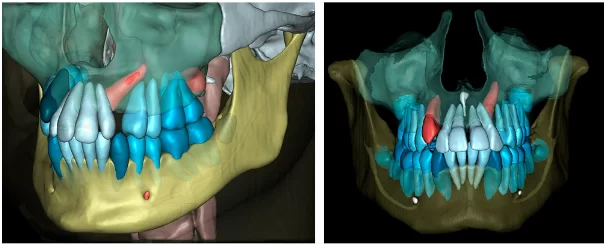
As a specific example, let’s consider the segmentation function of the popular Diagnocat program, specifically the function based on cone beam computed tomography (CBCT) data. Manual assembly of a 3D model requires high-level skills and the use of an expensive software package. And then the correction of defects caused by micromovements of the patient’s head, glare from metal elements in the patient’s jaws, and so on. The Diagnocat service automatically converts a series of CT scans into a 3D .STL file.
These files cannot always be used to develop custom prostheses without additional verification and correction. However, the data obtained makes communication between the dentist and the patient more informative, and for the specialist it simplifies the choice of treatment method and the selection of the necessary tools.
Let’s consider several examples of using the function of converting CT scans to a 3D file.
- Orthodontists often find it difficult to explain why certain procedures are necessary. The use of abstract models is not always accessible enough for the patient. And a 3D model of the patient’s own jaws and face is very clear. Moreover, in the program environment, you can change the transparency of soft tissues and clearly demonstrate how the pathology looks, and what specific steps will be required for treatment, and justify the necessity and cost of treatment.
- Surgeons often need to create a surgical template for precise implant positioning. This usually requires intraoral scanning data and cone beam computed tomography (CBCT) data, followed by processing the resulting files and developing the template on third-party software. Combining data from different scanning methods and linking the model into a single coordinate system was a challenging task. However, in the development environment of the Diagnocat program, it is possible to combine data from CBCT and intraoral scanner into a single coordinate system and develop an individual surgical template. This speeds up the process several times.
This is only a small part of the practical benefits. In addition, a 3D model of the patient’s skull and jaws helps to develop aligners for orthodontic treatment, assess the volume of bone tissue deficiency and justify the need for bone tissue augmentation for implant placement, and many other useful functions.
We hope this news was useful for you and our readers will get practical benefits.


