Synthetic Materials for Fillings Could Soon Be a Thing of the Past: Scientists Have Developed a Successful Model for Restoring Biological Enamel
In the realm of modern dentistry, the treatment of cavities and enamel damage has long relied on synthetic white filling materials. However, a groundbreaking breakthrough on the horizon is poised to reshape this landscape entirely. Researchers at KU Leuven, led by the esteemed Professor Hugo Vankelecom, in collaboration with Professor Annelies Bronckaers from UHasselt, have unveiled a pioneering 3D model featuring human dental stem cells. This innovative research, recently published in the prestigious Cellular and Molecular Life Sciences journal, could potentially revolutionize the way we approach dental restoration.

Delving into the Cutting-Edge 3D Dental Stem Cell Model
Our teeth play a vital role in various facets of our lives, from the basic acts of eating and speaking to our emotional well-being and self-esteem. However, the intricate nature of human teeth has presented challenges in the realm of research. Unlike rodent dental stem cells, certain human dental stem cells have proven challenging to cultivate in laboratory settings. This very obstacle prompted the ingenious development of the 3D research model by the KU Leuven and UHasselt collaboration. This model incorporates stem cells derived from the dental follicle, a specialized membrane enveloping unerupted human teeth.

 In Tooth Organoid Medium (Tom) (Passage 0, P0), And Robust Passageability (Brightfield Pictures Of Indicated P). B Histological (H&Amp;E) And Ultrastructural (Tem) Analyses Of Tooth Organoids Grown In Tom For 14 Days. Box And Arrow Indicate Cuboidal Epithelium (Ce) And Squamous Epithelium (Se), Respectively. C-E Brightfield Phase-Contrast Images And Immunofluorescence Staining Pictures For Markers As Indicated, Of Primary Df Tissue And Full-Grown (Day-14) Organoids. Arrows Indicate Double-Positive Cells Of Indicated Markers. Boxed Areas Are Enlarged. Dapi (Blue) Was Used To Label Nuclei. Scale Bars: 50 Μm, Unless Indicated Otherwise")
Establishment of organoids from human dental follicle. a Schematic of organoid culture set-up. Progressing development of organoid structures after seeding dissociated dental follicle (DF) in tooth organoid medium (TOM) (passage 0, P0), and robust passageability (brightfield pictures of indicated P). b Histological (H&E) and ultrastructural (TEM) analyses of tooth organoids grown in TOM for 14 days. Box and arrow indicate cuboidal epithelium (CE) and squamous epithelium (SE), respectively. c-e Brightfield phase-contrast images and immunofluorescence staining pictures for markers as indicated, of primary DF tissue and full-grown (day-14) organoids. Arrows indicate double-positive cells of indicated markers. Boxed areas are enlarged. DAPI (blue) was used to label nuclei. Scale bars: 50 µm, unless indicated otherwise
Unveiling the Unique Advantages
Professor Vankelecom emphasizes the distinctive advantages offered by this groundbreaking 3D model. By accurately replicating the original properties of stem cells, this model essentially recreates a miniature segment of the human body for research purposes. The utilization of dental stem cells enables the creation of various dental cell types, including ameloblasts—crucial contributors to enamel formation.
Pioneering Biological Enamel: A Paradigm Shift in Dental Restoration
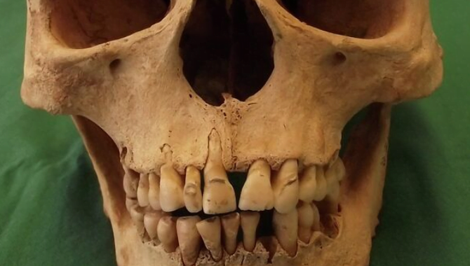
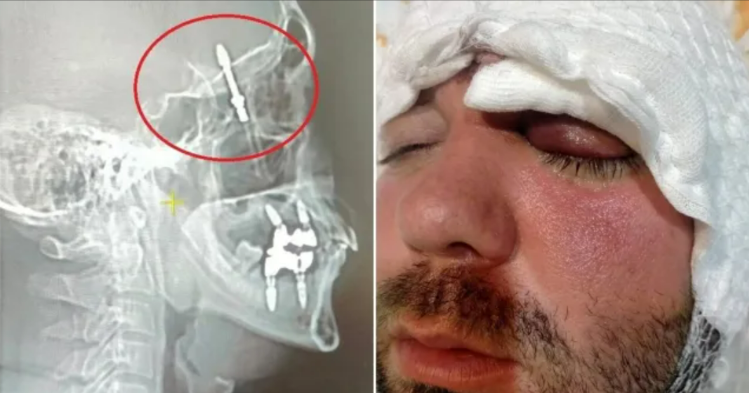
The daily exposure of our teeth to acidic and sugary foods makes them susceptible to enamel damage, necessitating immediate dental intervention. Historically, dentists have relied on synthetic materials for treating cavities and enamel erosion, which stand out as foreign objects in the X-ray below. However, the limitations of such materials have paved the way for a monumental breakthrough.

X-ray with synthetic fillings
From Stem Cells to Enamel Components
In this remarkable 3D model, dental stem cells undergo a transformative process, maturing into ameloblasts—cells responsible for producing enamel components. This monumental advancement holds the potential to yield biological enamel, a natural alternative for repairing dental enamel. Unlike synthetic materials, this biological enamel not only naturally restores the physiology and functionality of dental tissue but also minimizes the risk of complications like tooth necrosis.
Multi-Faceted Impact: Beyond Dentistry
The implications of this pioneering research extend far beyond the realm of dentistry. The versatile 3D cell model offers applications across various sectors, heralding a new era of research and innovation.
Unveiling Insights in the Food Industry
One notable application resides within the food industry. The 3D cell model could serve as a valuable tool for assessing the impact of specific food products on dental enamel. By offering insights into the effects of different consumables on oral health, this breakthrough has the potential to steer the food industry towards more enamel-friendly offerings.
Elevating Oral Care Products
Furthermore, toothpaste manufacturers stand to benefit from this paradigm shift. The 3D model’s capabilities could be harnessed to optimize the protective and restorative qualities of oral care products. This innovation could lead to the development of toothpaste formulations that are exceptionally effective in safeguarding dental enamel.
Charting a Course for the Future: A Blueprint for Dental Innovation
Professor Vankelecom’s aspirations extend even further. While the current focus lies on ameloblasts, the newfound potential of this 3D cell model opens doors to a myriad of possibilities. The amalgamation of various dental stem cell types could pave the way for the creation of diverse tooth structures, and perhaps, an entirely biological tooth.
In conclusion, the groundbreaking 3D dental stem cell model developed by the collaborative efforts of KU Leuven and UHasselt holds immense promise for the future of dental restoration. As research continues to unravel the full extent of its capabilities, it is evident that this innovation has the potential to reshape dentistry, impact various industries, and ultimately enhance the quality of life for individuals worldwide. The journey towards a natural, effective, and revolutionary approach to enamel repair has begun, and the world of dentistry stands on the cusp of transformation.