Contents
We continue the series of articles about the history of dentistry. In the first part we took apart the times from the ancient world and antiquity to the Renaissance. In the second part, we will look at an equally interesting period, namely the 19th and 20th centuries. It was then that fundamental discoveries were made and surgical techniques and protocols were developed that are still in use today.
Implantation and dental treatment in the 19th century
The early 19th century is interesting for us because in 1809, a book by Dr. Maggiolo, an Italian by birth, was published, describing the method of dental implant manufacturing and installation, which were placed in the cavity of a tooth that had just been extracted.
There were many things described in the book about dental treatment techniques, but we are interested in the first described protocol for making and placing complete heterogeneous implants.
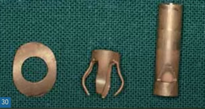
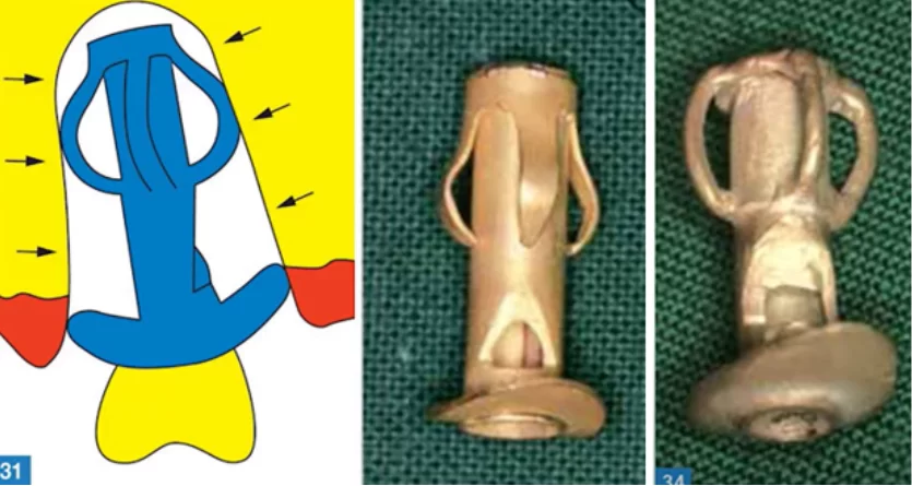
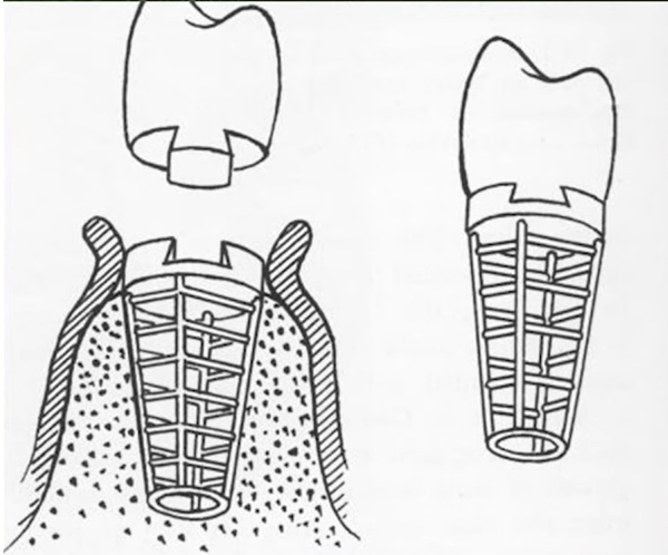
The implant was made from a gold tube to which a part with petals was soldered, as well as a flat skirt in the coronal part of the implant, see illustration below.
Dr Maggiolo described the tooth extraction procedure in detail and insisted that it be as atraumatic as possible. He described how bone ingrowth into the petals and other cavities in this implant would be expected. One month later, he recommended a crown to be placed on this implant. And he described in detail the process of making this post crown.
This work made a great impression on his contemporaries. Some specialists cited it, while others objected. It is known that 46 years after the publication, the English medical scientist criticized Maggiolo’s method. He tried several times to insert an implant according to the described protocol and received a number of serious complications.
But there were many successful implantations done by Dr. Maggiolo himself and his followers. The doctor himself considered his methodology flawless and did not accept criticism, but his followers agreed that not everything was as perfect as they would have liked.
Patients complained of:
- inflammation of the gum around the implant;
- unpleasant odor, etc.
And there is an explanation for all this, because the fit of the tube was not perfect and biological fluids with microbiota got into the implant cavity. In fact, there were quite a few problems and today we can reliably say why there were so many complications and limitations of this technique:
- The use of a gold alloy with a lot of impurities. 18 carat gold was used, which contains cytotoxic materials such as copper, silver and some others. And even more toxic impurities contained gold brazing compound. The purer 24 karat gold is biologically neutral and would have been better suited for implants.
- Lack of effective anesthetics, antiseptics and anti-inflammatory drugs at the time. As well as a culture of using already known medications in dentistry.
- Inability to monitor the insertion and the process of osseointegration. This is now done with X-rays.
- Implants were only suitable for immediate placement after a tooth extraction. And only for single root teeth. It is impossible to place such a prosthesis in place of a molar.
Nevertheless, it is in Dr. Maggiolo’s work that we first encounter:
- With the documented principle of osteogenesis. He founded that the implant would gain stability through the growth of new bone in the cavities of the metal structure.
- The principle of two-stage implantation. The first stage is the integration of the implant through bonefilling. Only after the integration process is complete, a post crown is placed and the implant is then placed under load.
- Finding primary stability during implant placement as a prerequisite for osteogenesis.
The nineteenth century was a breakthrough in many areas and one would like to give credit to non-dentists who in one way or another influenced the development of medicine in general and dentistry in particular. First of all, the discoveries concerned asepsis and anesthesia.
- Nikolai Ivanovich Pirogov (1810-1881), who in 1844 guessed that the cleanliness of the operating field as well as the aseptic preparation of personnel and instruments directly influenced the prognosis of inflammation after surgery. It is noteworthy that it was just a guess, because Pirogov could not prove his hypothesis in any way, there were still more than 10 years before Louis Pasteur’s discovery.
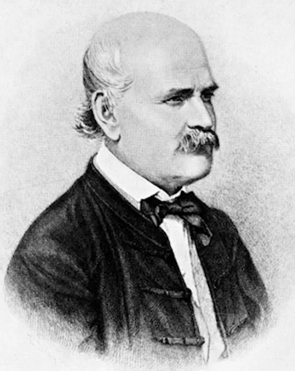
- Ignatz Philipp Zemmelweis (1818-1865) – saved an incredible number of women in childbirth thanks to his genius insight into the introduction of asepsis into obstetrical procedures. Before that time, a great number of women in labor had died of so-called maternal fever. In most cases, the midwives of the time, and especially the students, interacted with women in labor without even washing their hands after working with cadavers. Unfortunately, the fate of this talented scientist is tragic. Unfortunately, his authority was not as great as Pirogov’s, and during his lifetime he was severely criticized, persecuted and even placed in a psychiatric hospital, where he died at a very young age. Today in Budapest there is a monument to Zemmelweiss with the inscription: “Savior of Mothers.
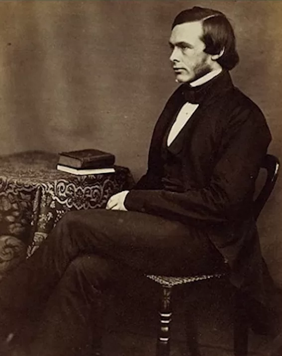
- Joseph Lister (1827-1912) entered the history of medicine through his 1867 monograph in which he described surgical protocol in detail. He described in great detail how to treat the operating room, disinfect instruments, aseptic treatment of surgeons’ and assistants’ hands with carbolic acid. He treated absolutely everything with it and even sprayed it in the operating room with an aerosol. This was not very good for the health of doctors and assistants, but it was extremely beneficial for the success of patient survival. It is thanks to his personal experience and his published monograph that Lister is rightfully considered the father of the founder of modern aseptic and antiseptic techniques.
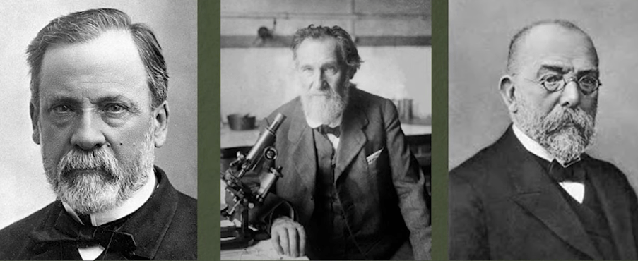
- The great trio of scientists who made key discoveries in microbiology:
– Louis Pasteur (1822-1895).
– Ilya Ilyich Mechnikov (1845-1916)
– Robert Koch (1843-1910).
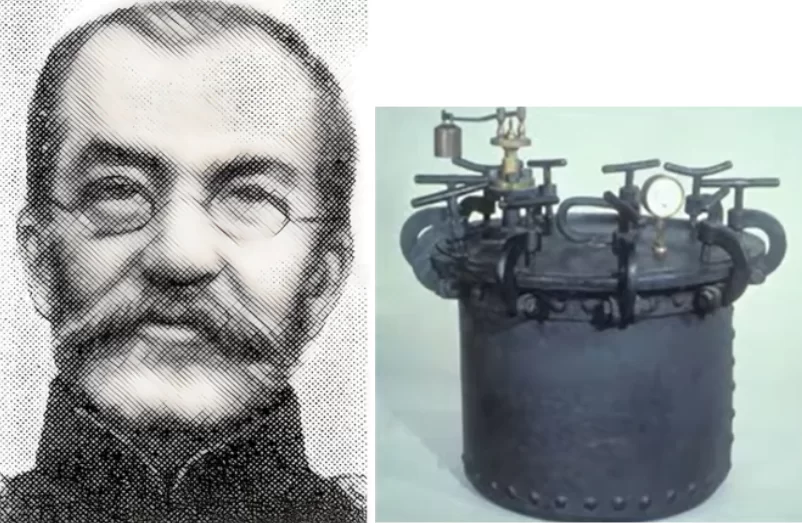
They made a tremendous contribution to the concepts of pathogens in the workings of the immune system. They discovered the causative agents of tuberculosis, rabies and anthrax. It was their discoveries that provided a theoretical basis for the conjectures of their predecessors, and their practical recommendations made asepsis and antisepsis mandatory in medicine. - Ludwig Heidenreich (1846-1920) was the first to use the autoclave to sterilize instruments and published a paper in which he pointed out the importance of pressurized steam treatment of surgical instruments.
More fundamental discoveries were made in the nineteenth century than in any other before or since. Next we will look at the chemicals that came into use in surgical operations in general and dental operations in particular.
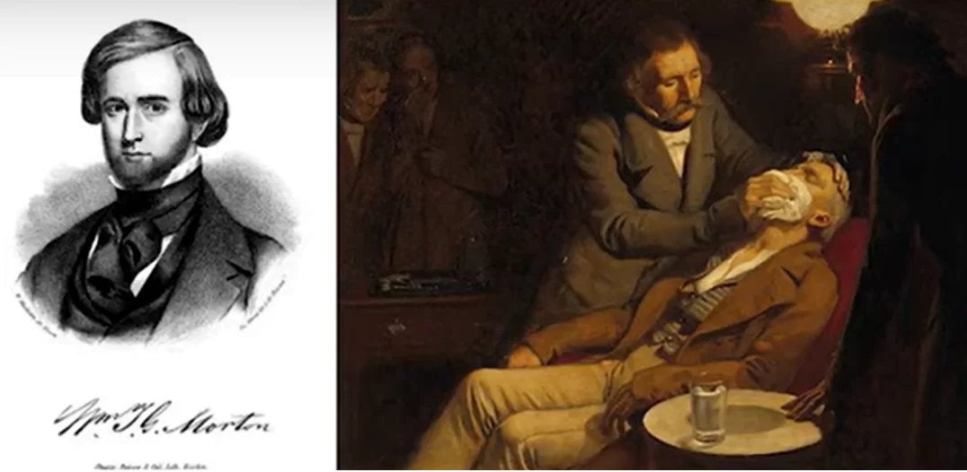
We will begin with the production of ether and then chloroform, which for a long time were used to immerse the patient in a state of anesthesia. And the first person in the world to use ether anesthesia was Dr. William Thomas Green Morton (1819-1868). He did it in a clinic in Boston in 1846 and made a detailed description of the operation.
Morton was removing a mandibular tumor from a patient, and the operation was successful. Less than a year later, etheric anesthesia became actively used in Europe and Russia.
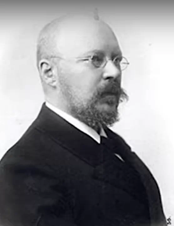
The middle of the 19th century can be considered the moment of appearance of effective anesthesia, which opened new possibilities for various reconstructive surgeries. In connection with the research of anesthesia methods, it is worth mentioning Vasily Konstantinovich von Anrep (1852-1927). His discovery is not specialized, he was more concerned with pharmacology and was not a practicing surgeon. Nevertheless, it was he who published an article describing the use of cocaine as a local anesthetic. He injected it subcutaneously and got good results.
As early as the 20th century, novocaine, lidocaine, and the modern standard of local anesthesia, articaine, appeared. But these are all derivatives of 19th century discoveries, because it was then that it became clear that it was possible to block pain receptors locally.
It was these discoveries that made it possible to perform complex surgeries where you have to drill into the bone, shape the bed, that is, operate more freely to be resourceful.
And in this light, we will talk about doctors, some of whom lived in the 19th century and some of whom continued to practice in the early 20th.
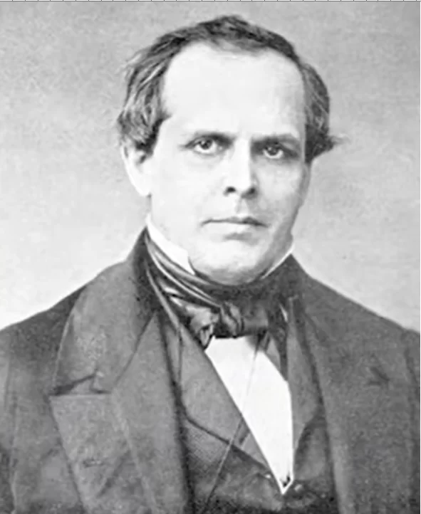
- First we must be sure to mention Dr. Chapin A. Harris (1806-1860) who famously, along with his colleague Dr. Hayden, created one of the first dental schools (faculties) in Baltimore. Dr. Harris created his implant system based on a lead-coated platinum implant. It was well known at the time that lead was not susceptible to corrosion. And scientists at the time hoped that lead would protect the implant reliably and be biologically compatible. In fact, lead is quite a toxic metal. But nevertheless, the method of implantation has already been described in detail, it was associated with the formation of the bed and installation of the implant under sterile conditions with the use of anesthesia.
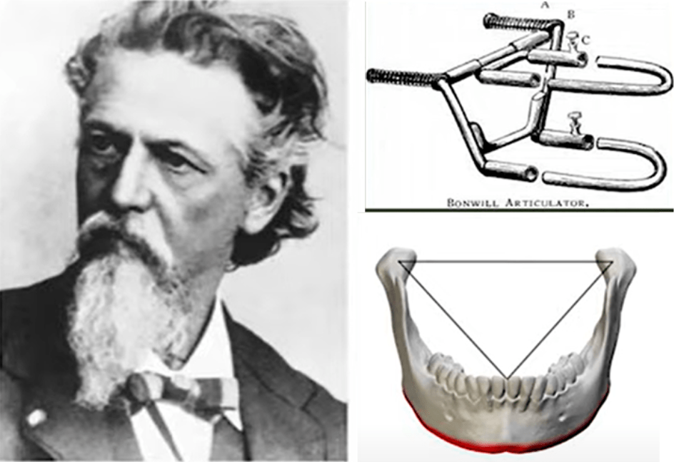
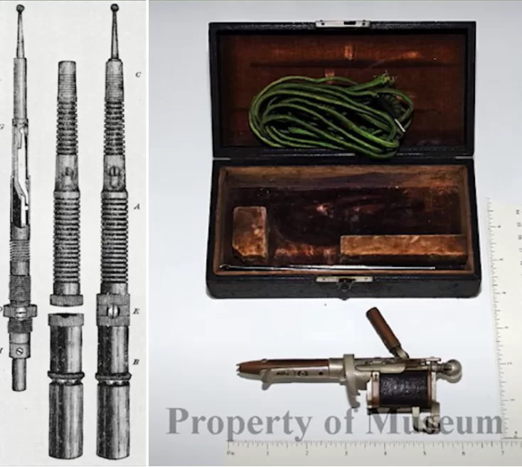
- William Bonwill (1833-1899), well known to all dentists by the Bonwill triangle, was probably the last specialist whose entire body of work was created in the 19th century. He was the inventor of the first and in fact still in use today, the Bonwill articulator is shown in the illustration below. This apparatus simulated jaw motion and allowed for the design of dentures based on the alignment of the teeth according to jaw movements.
He was both a scientist, physician, and engineer, and received many patents during his lifetime. In particular, he developed a system of handpieces, was the first to use an electric motor as a drive for drilling. He invented the drill system and this all allowed him to create his own system of implants made of gold and iridium alloy, which allowed him to increase the strength.
He made implants with complex configurations with holes and a porous surface for bone ingrowth.
Development of Implantologists in the First Half of the 20th Century
We have reached the 20th century, which was spent searching for and creating the perfect implant system. By the beginning of the 20th century a sufficient theoretical basis and practical experience had already been accumulated. We will not be able to list all the researchers so let’s go over the most important discoveries and developments that determined the future fate of implantologists.
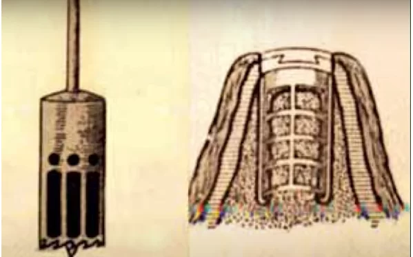
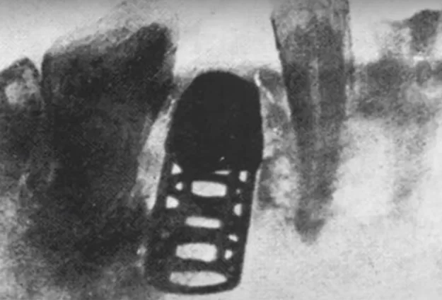
- Dr. EJ Greenfield’s basket implant – 1913. This system really resembled a basket made of platinum-iridium alloy and was installed with a special drill resembling a modern trepan, see illustration below.
Emphasis was placed on intraoperative antisepsis and precise preparation of the implant site. Greenfield’s innovation was also the dovetail wedge connection between the crown and the abutment. Dr. Greenfield lived at a time when radiography was already in active use and evidence of successful integration of implants of his design has come down to us.
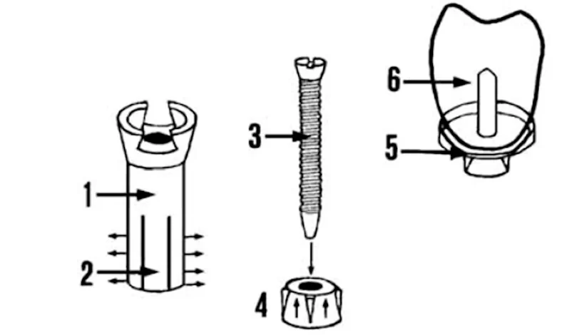
2. The first screw fixation suprastructure by Dr. Leger-Dorez (1920 France). In the illustration below you see a tubular implant with petals somewhat similar to the Maggiollo system.
The screw was supposed to lift the tapered nut, which was located in the area of the lobes and thus increase the stability of the structure. In fact, this implant system was not very successful due to the large number of complications. Now we understand why, according to protocol, the screw had to be screwed in after healing and partial or complete integration of the implant. The movement of the petals traumatized the bone tissues, causing a lot of inflammation and frequent loss of implants.
3. Dr. P. B. Adams (1937) implant system where a cylindrical ribbed implant with a screw-on ball attachment was used to secure a removable denture, see illustration below.
This was an innovative two-stage restorative system with plugs and gingival shapers, the design of the Adams system is very similar to modern systems.
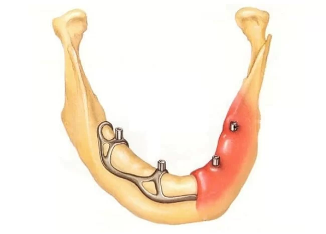
4. Dr. Dahl’s subcutaneous implant system (1940 Sweden). He used stainless steel for this system was designed for the restoration of the mandibular dentition. And this system had quite good indicators for that time:
- 10-year survival rate – 60%;
- Survival rate after 20 years was 40%.
This design was not very popular in Dr. Dahl’s homeland, due to the conservative nature of the Swedish school of dentistry, but the enterprising Americans Golden and Gershkevich have adopted this technology and quite successfully. So much so that these doctors founded the American Dental Association, which still exists today.
The second half of the 20th century and today – the Branemark era
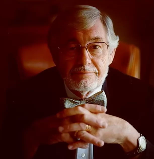
This part will focus on just one person – that’s Per-Ingvar Branemark (1929-2014). This Swedish scientist made a defining contribution to modern implantology. Immediately after graduating from Lund University, Branemark became a practicing physician. It was there that he began work on his dissertation. There he also worked in the laboratory of vital microscopy, all this taking place at the end of the 50s.
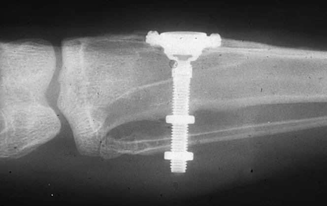
There he studied the reaction to trauma and bone marrow healing and proved that bone tissue is alive and needs to be treated with care. He wanted to study the microcirculation of blood in bone marrow tissue and its change during the healing process. To do this, he needed to place a tube in the tibia of a rabbit that would stand for an extended period of time. This was necessary in order to continuously take microphotographic pictures of the changes for subsequent histological and visual analysis.
Branemark was greatly assisted in his research by his mentor, Professor Hans Emneus, who recommended that he make the tube from titanium. At that time, titanium as a material for biocompatible structures was already in use and considered quite promising.
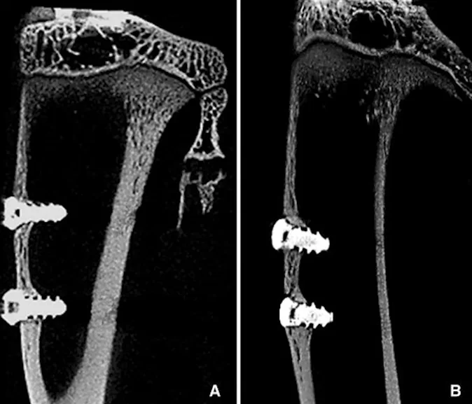
But we are interested in the end of the experiment, when Branemark tried to remove these tubes, through which images were taken from the animal bone. It turned out that the tubes were very firmly fused to the bone and almost impossible to remove. Another researcher would have paid little attention to this and simply disposed of the rabbit and removed the valuable material by destroying the bone. But Branemark decided to study this phenomenon separately, even though it was not the purpose of the study.
Branemark made slices and conducted a histological study and found that bone tissue sprouted into all the irregularities and roughnesses on the surface of the titanium parts. That is, there was attachment of bone tissue to the titanium tubes.
Branemark discovered this effect in 1956, and in 1960 he moved to Gothenburg, where he became professor of anatomy. At the same time, he continued his laboratory research of his discovery. He studied the behavior of titanium in different bone structures and conducted experiments on dogs and pigs, etc.
He was gathering like-minded people around him. At that time he was joined by two most interesting people. The first was Victor Kuijka, a watchmaker, who initially made all the parts for the research and the first Branemark implants. He knew how to work with metal and make parts with the highest precision. And the other Richard Skalock is a bioengineer and he knew very well how metals behave in the body and he was very helpful in the research. And this group, driven by Branemark’s enthusiasm, was accumulating more and more data, and eventually there came a time when it was necessary to move from animals to human testing.
It should be noted that by that time titanium was already being used in the human body, especially by orthopedists and traumatologists for fixing broken bones. But for a long time Branemark could not find a patient for his experiment, until he met a man named Gosta Larsen. His name is written in the history of medicine because of his serious problems. This man had been disabled since childhood and by the age of 34 he had a cleft lip, cleft palate, had undergone several surgeries, and was hearing impaired. But most importantly in this context, he was practically unable to chew his food properly because the removable dentures weren’t holding up well. And by that time he had full adentia.
Specialists understand how difficult the task of prosthetics is for a patient with a cleft palate. It was he who heard from his attending physician about Branemark’s experiments. And he approached him himself. Branemark originally had no plans to use titanium and the osseointegration phenomenon he had discovered in dentistry, but Gosta Larsen was in the right place at the right time. He had four implants placed on his lower jaw with a fixed suprastructure. And that changed this patient’s life dramatically, he was able to chew normally. He was an enthusiastic fan of Branemark and followed him for a long time. And one day on his way back from an examination, he enthusiastically told the cab driver about such a wonderful doctor who had performed a miracle. And so it was a coincidence that the cab driver was his peer, he was also about 35 years old, and also toothless. The cab driver’s name was Sven Johansson, and he became Dr. Branemark’s second patient. This, too, was a very successful case of dental restoration with titanium implants. At the time of writing, Sven Johansson is alive and still using his dentures.
It was with these two patients that the Branemark implant system began such tremendous success. After the initial successes, Branemark began to actively promote his system; he set up a small company manufacturing implants and implant accessories. He tried to get as many doctors as possible acquainted with his development. But unfortunately, he was not well received at first, due to the conservative nature of the Swedish medical community.
It got so far that in 1974, a collective complaint was filed against Branemark with the local Ministry of Health, demanding an investigation to stop the activities of the charlatan. The Swedish Ministry of Health did not forbid it, but requested an experiment in which 20 patients were chosen for whom Branemark implants were placed and in 1975 it was confirmed that the method was completely reliable and very promising. And it led to the fact that in 1976, Branemark received official confirmation from the local counterpart of the FDA that the use of titanium implants Branemark is safe and recommended for use.
During his long life, Branemark alone and together with his colleagues published 168 papers not only on dentures and osseointegration but also on other fields of medicine. Many works were devoted to the study of the bone marrow microcirculation, the influence of drugs on the regenerative processes in bone tissues, etc.
But back to the development and distribution of titanium implants of Branemark design. The breakthrough came in the late ’70s, when Professor George Zarp learned about Branemark. He was in the Department of Orthopaedics in Toronto. He was a renowned specialist who had begun practice as early as 1966. George Zarp even flew to Branemark in Sweden, then repeated the same experiment in Toronto with 20 patients, and the results were just as successful. It turned out that Branemark’s enthusiasm was supplemented by Zarp’s energy. It was Zarp who convinced Branemark to attend the Toronto conference. This is the famous 1982 conference from which modern dentistry quietly began its countdown. More precisely, the mass spread of titanium implants. Branemark’s students went around the world and started their own practice, new ones arrived in their place, most of the conference participants, who were already experienced specialists, started to use the titanium implants offered by Branemark.
The key feature, even the basis of Branemark’s discovery was a clear understanding of the processes of osseointegration, how bone tissue behaves in the healing process, he clearly described how osteocytes are attached to the implant surface and what to do to make osseointegration as good as possible.
Branemark emphasized that bone tissue is a living structure and must be treated with the utmost care. It’s not that people didn’t understand this before, but even doctors treated bone as something like wood, with which they could do anything, drill, screw in pins, etc. without much concern about what would happen in the long run.
Branemark developed and defined the optimal clinical protocol for implantation:
- implant placement with full flap suturing for four months on the lower jaw and six months on the upper jaw;
- After the required time, the soft tissues were opened and a gingiva former was placed in the implant;
- Approximately one month later, impressions were taken and the substructure of the permanent denture was fabricated.
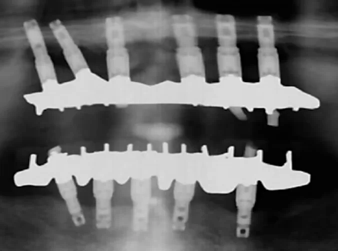
For complete edentulism, Branemark recommended placing four implants on the lower jaw and six on the upper jaw and attaching fixed dental restorations to them. Later, his followers expanded the indications for implantation, modifying the protocol depending on the clinical picture of the patient, but Branemark’s development was the basis.
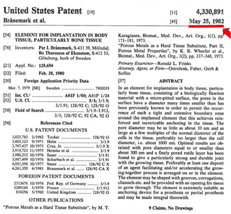
Branemark filed an application in 1979 and received a patent for his design in 1982. Note that Branemark’s patent contains no pictures or diagrams of his implant.
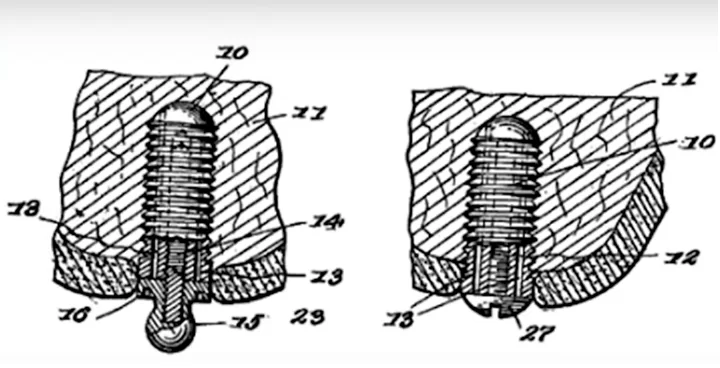
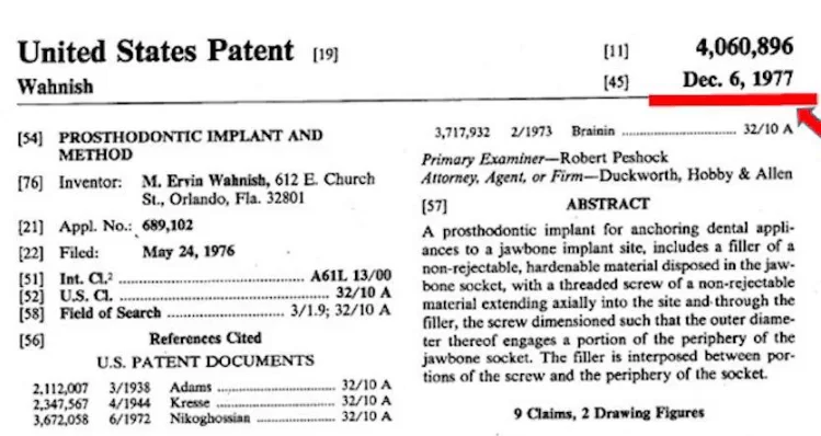
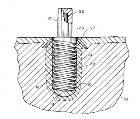
In fact, the design of the root implant with thread coils for bone contact and screw-retained suprastructure was invented before Branemark, see the 1977 patent.
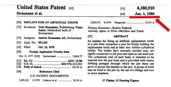
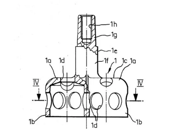
Moreover, just a couple of years before Branemark, Dr. Straumann received a patent for a titanium implant, which he developed together with Dr. Schroeder. But that’s a whole other story, which we’ll probably cover in a separate article.
Many people wonder why Branemark was not awarded the Nobel Prize, since his contribution to medicine was more than significant. But the Royal Swedish Academy decided otherwise. The fact is that in 2004 the Belgian University and several other famous scientists applied for the Nobel Prize for Branemark, but it was rejected. And technically the Royal Swedish Academy was right. They analyzed the scientific works of Branemark himself, studied the works of other specialists in this field and discovered that long before Branemark there had been other works devoted to the use of titanium in medicine and the integration of bone tissues with the surface of titanium. The spiral threaded root implant was also invented before Branemark, as evidenced by the patents on root implants, some of which were also made of titanium.
Technically, Branemark really had no primacy in any field. His main merit was overcoming the inertia and conservatism of the medical community. Branemark literally forced the whole world to adopt new standards. So in terms of the size of his contribution to humanity, Branemark is well worthy of the Nobel Prize.
This can be clearly seen in the difference in the results of the 1978 and 1982 conciliation conferences of dentists.
In 1978, a consensus conference on dental implantology was held at Harvard, where unified standards and norms were adopted for what counted as success in the treatment of adentia. Scientists and practicing dentists recorded the following as the norm:
- Five-year survival rate of 75% or greater;
- Mobility of the implant is less than 1 mm;
- Bone resorption of not more than 1/3 of the implant length.
Looking at these indicators today it seems that this is a stupid joke and the license should be revoked for such indicators. Today it is hard to imagine a situation where bone resorption exposes a third of the implant. And the actual ten-year implant survival rate was less than 50%.
And just four years after the Toronto conference with Branemark, a new conciliation conference was held in which both Branemark and Professor Zarp participated. The results led to the adoption of new standards for implant and restorative success, which were later supplemented by Dr. Albrektson et al. in 1986:
- A five-year survival rate of at least 95%;
- ten-year survival rate of 80% or more;
- Complete absence of implant mobility;
- little resorption (less than 2 mm in the first year and less than 0.2 mm thereafter);
- absence of pain, inflammation, neuropathy, paresthesias, integrity of important anatomical structures;
- absence of bone dissection on the radiograph;
- adequate and functional dental restoration from the perspective of the patient and the dentist.
These norms were no longer very different from those of today and were quite consistent with the actual statistics of the surgeries that began to be done at that time.
It is this amazing leap in just four years that characterizes the contribution of Dr. Branemark and his followers to the development of dental implantology.
Of course, we could not do justice to all the scientists and practicing dentists who contributed to the development of treatment techniques. But we hope you found this article interesting and we will continue to bring you up to date on current discoveries and techniques.

