CAD/CAM Compatible with Paltop®
No products were found matching your selection.
CAD/CAM Technology and Prosthetic Components
In this section you can buy Scan Body markers, implant analogues, fixation screws as well as t-base screw-retained abutments for single restorations with delivery. Modern technology makes treatment easier, especially with regard to patient discomfort. Dentists and dental technicians are also finding it much easier to make even complex restorations without the numerous fitting and adjustment steps. Let’s take a closer look at the technology and the range of elements for CAD/CAM technology from Uniqa Dental.
CAD/CAM advantages and specific applications in dentistry
3D scanners, 3D printers and digitally controlled machines for the milling of dental prostheses have been actively used in dentistry since the early 2000s. The principles behind CAD/CAM technology are:
- Imaging and transfer to a computer file of the patient’s teeth and surrounding tissues. An intraoral 3D scanner is used for the imaging process, which takes a series of images at high speed. From a series of images from different positions, the software generates a three-dimensional image of the jaw with the teeth. Scanning heads are used to get a clear picture of what is happening in the areas where there are no teeth and dentures are to be placed. At the time of scanning, the dental implants must already be in place and osseointegrated. The gum shapers are removed and the heads (Scan Body) are put in place before the scan. Their position allows you to see the position and angles of the implants relative to the dentition. The installation of scan markers and imaging is also possible immediately after implant placement if immediate restorations are planned. A temporary resin denture is often used for immediate restorations. Temporary restorations are also fabricated using CAD/CAM technology, such as milled on a machine or printed on a 3D printer.
- Based on the generated 3D model, a physical model of the jaw is printed, on which complex restorations are tried and adjusted. In relatively simple cases, this step can be dispensed with and the denture can be immediately milled and fitted in place.
- In the fabrication of the denture itself, CAD/CAM technology can be used to fabricate the prosthesis:
- Finished prosthesis: Zirconia crown; a tooth row fragment with finished crowns or a complete dental arch for all four or all six dentures;
- A bridge bar on which the restoration will be placed;
- Other orthodontic constructions.
Actuators can be used:
- Milling machines with three axes of motion and digital control are capable of turning out very complex structures using the hardest materials;
- 3D printers for special purposes, after the model needs sintering (firing) to acquire the final strength.
Finishing includes polishing, placement of multi-unit sleeves in the denture or t-base abutments if the screw fixation is planned. The final step is the insertion of the denture into the patient’s mouth.
Our video showcases the digital dental procedure for screw retained implant-supported restorations, from dental implant placement to final denture placement.

A brief list of the advantages of CAD/CAM technology:
- Accuracy: Digital scanning does not depend on shrinkage or random deformation of the impression material. Modern scanners have a resolution in the range of 100-150 microns, and this significantly exceeds the reliability of a digital model compared to that obtained with an impression.
- One-step production of highly complex structures with an ideal fit.
- Comfort for the patient: Taking an impression is a long and extremely uncomfortable process, which makes many patients gag. Whereas scanning with an intraoral scanner does not take long and does not cause noticeable discomfort.
- Speed of production: If all necessary equipment is available and ready to work, the time from the creation of the 3D model to the readiness of the dental prosthesis is about one hour. In the case of complex operations, of course, more time is required, but still several times quicker compared to traditional methods. There is no need for numerous fitting procedures, etc.
- The prosthesis design file is stored in the clinic’s archive. If necessary, a copy of the prosthesis can be made as quickly as possible.
- Absence of internal overstresses and complications due to inaccurate fit of the denture. Passive fit is ensured by the high precision of the 3D model as well as the accuracy of the finished restoration.
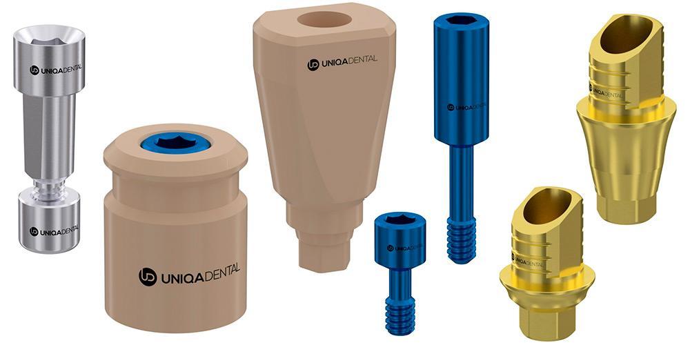
Implant analogues from Uniqa Dental for CAD/CAM technology
For modeling and fitting bridge structures, our product range includes analogues of implants. Namely:
- A digital analog of the implant with a standard hex interface. It can be fitted with any abutment that is compatible in terms of attachment. The implant analog is a reusable product. The titanium implant analogue is placed in the 3D model of the jaw with high precision. This allows the dental technician to calculate and fabricate a single crown or crown restoration with minimal deviations. Fitting and adjustment, if necessary, is very easy. A printed model of the jaw much more accurately conveys all the anatomical features compared to the impression that was created with the help of transfers. The finished denture design is transferred to the dentist and fitted to the patient with a good passive fit without the need for additional manipulation. It is important to correctly place the abutment analogue in the model, for which the 3D printer must be properly calibrated. Uniqa Dental provides all the necessary electronic files to set up and calibrate the equipment.
- Analogue of the multi-unit abutment: More specifically, a single part that mimics an implant with a screw-retained abutment already in place. This saves money, because you need to buy just one part instead of the analog of the implant and the abutment. The principle of working with a digital analogue MUA is similar to the previous option; the key difference will be that you need several such analog MUAs for CAD/CAM. Because the multi-nit system is only used for group dental restorations, they are placed in the printed model according to the actual placement of the implants in the patient’s mouth.
Scan markers (Scan Body) from Uniqa Dental
The most important task for the creation of a restoration is to obtain a model of the patient’s jaw with the exact location of the teeth and implants placed.
In the past, a physical impression had to be taken with transfers. Then you had to wait for the plastic compound to harden and take the impression, which was very uncomfortable for the patient. Now you can create a digital file with an intraoral scanner. This device takes a series of high-frequency images from which a 3D file is generated. While the teeth and soft tissues are clear, there will be gaps where the missing teeth were. Such a model is of no use for the creation of a prosthesis. That is why scan markers are used to mark the locations and angles of the implants.
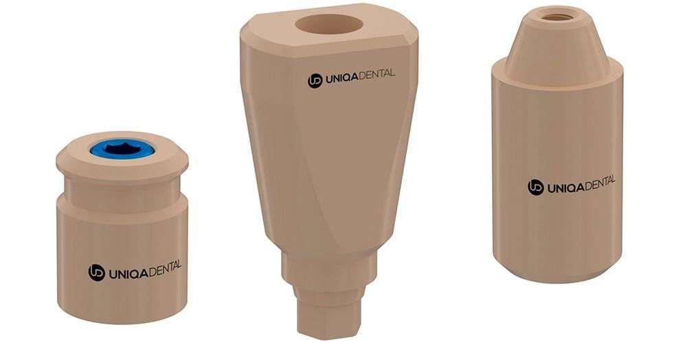
In this section you can buy scan markers in several designs from Uniqa Dental. They are made in such a way that they fit into the frame and have a good appearance when scanned with an intraoral scanner. Uniqa Dental scan abutments have the following characteristics and varieties:
- Made from PEEK plastic that is strong enough to withstand repeated use (at least 20 times).
- The surface is made matt to show better when scanning. There are some scan abutments made of titanium, and to keep the surface from shining, it is either roughened or coated with a special spray. Plastic, however, is much easier to use and cheaper, so we suggest buying scanners made of PEEK plastic.
- You can buy scanned abutments of cylindrical shape with an interface for screw fixation MUA D-type. These are placed on top of the abutment already inserted in the implant. The height of the scan marker is, therefore, often shorter compared to the scan marker that is inserted into the implant. There are, however, some occasions when greater heights are needed, so you can buy a scan body marker up to 12 mm in height. In addition, you can buy screws to fix the scan marker to the abutment.
- Scan markers with installation directly into the implant: This is a more common variety and allows determining the position and angle of the implant thanks to its blade shape. Using the scan and 3D model, a customized abutment can be selected or fabricated, which is indispensable in complicated cases. Standard abutment screws are used to attach the scan marker to the implant. Since this is a temporary construction, it is cheaper to use laboratory screws rather than clinical screws.
T-base abutments for screw-retained single crowns
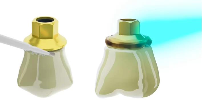
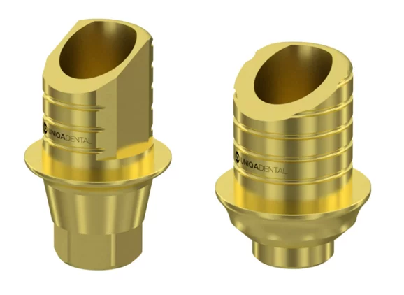
Abutments with screw-retained prosthesis are used in conjunction with the CAD/CAM system and rarely with cemented retention. T-base abutments for single implants can be purchased in this section. Screw-retained crowns have a number of advantages over cemented retention. Let’s look at these in more detail:
- Ease of placement for the dentist: All operations with adhesives and cements take place outside the patient’s oral cavity. This means it is convenient for the specialist to grind and remove any cement residue that may protrude at the junction between the crown and abutment.

Even a small amount of cement that comes into contact with the soft tissue of the gum can cause irritation and chronic inflammation. Of course, in most cases the dentist manages to remove cement residue from the patient’s mouth as well. However, this process is long, labor-intensive and does not provide a 100% guarantee in contrast to a screw-retained platform. In addition to the removal of cement, many other operations to create a crown occur outside of the patient’s mouth, which increases reliability and reduces the risk of errors and complications. After all, the dentist can work comfortably and see the smallest nuances of the process. - There is little or no micro-mobility in the implant/abutment line. The T-base abutment is placed with the crown already glued on it. The abutment with the finished restoration is tightened with considerable torque. After the t-base abutment has been screwed in, only one procedure remains: the closure of the screw shaft. The classical procedure, on the other hand, involves positioning the restoration on the abutment in the patient’s mouth. The screwed abutment is then subjected to considerable stresses that can loosen the connection between the implant and the abutment. If there is a gap and micro-mobility, then there is the so-called pump effect, when saliva containing bacteria is drawn into the internal structures of the soft gum tissue during chewing movements.
- If the crown has to be removed for any reason, the screw shaft must be drilled out to gain access and the t-base abutment must be removed along with the crown. Whereas with cement fixation the crown has to be physically destroyed. This is much more difficult and causes great discomfort to the patient.
T-base abutments from Uniqa Dental have anti-rotation notches on the surface, as well as slots on the side surfaces, which prevents the crown from turning relative to the titanium base.
Interface options are available for t-base abutments for a standard hex and for a 22° and 24° cone.
All Uniqa Dental prosthetic components are made of Grade 23 titanium-aluminum-vanadium alloy. This alloy was chosen because of its high strength values and is commensurate with the popular Grade 5 alloy, but surpasses it in terms of ductility (Young’s modulus), which is particularly important in the fabrication of thin components that work on fracture and torsion.
Another advantage of t-base abutments from Uniqa Dental is the precision manufacturing. We invest in our equipment and our specialized personnel. We, therefore, have minimum dimensional tolerances. Many dentists have had problems with dimensional inaccuracies. It is impossible to detect a difference of 50-100 microns by eye and the actual placement of such an abutment leads to a large enough gap. This results in micro-movement of the entire structure and the pap smearing effect that we have already mentioned.
You can order test samples of some components from us at a special price of $1. To get them, please leave an application with your address and contact details.


