Customizable Strontium-Enriched Scaffold May Improve Tissue Healing Around Implants – New Research Findings
A team of researchers from the University at Buffalo has developed a novel skeletal structure saturated with strontium, which can be personalized to fit any dental implant size. This innovation has the potential to enhance the healing process and tissue attachment in patients.
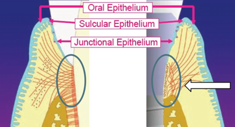
The Critical Importance of Connective Tissue Cuff for Implant Success
The success of dental implants relies on the growth and attachment of soft tissues to the implant surface. Previous studies conducted by the University at Buffalo researchers have shown that strontium, an element known to enhance bone density and strength, also supports the function of soft tissues. They discovered that strontium promotes the activity of fibroblasts, a type of cells responsible for forming connective tissues and playing a vital role in wound healing.
A dense ring of connective tissue should form around the implant. The quality and quantity of connective fibers are much better around a natural tooth. The goal of the research is to find a way to facilitate the rapid formation of a dense ring of collagen fibers around the implant. High-quality connective tissue attachment reduces the loss of bone tissue around the implant and minimizes the risk of peri-implantitis.
A recent study published earlier this year in the Journal of Biomedical Materials Research demonstrated that skeletal structures saturated with strontium, even at low concentrations, promote wound healing by stimulating the activity of gingival fibroblasts.
“Materials research for skeletal structures that enhance bone and skin healing has been conducted before, but adaptation for the oral cavity has been limited,” says lead researcher Michelle Visser, PhD, Assistant Professor in the Department of Oral Biology at the University at Buffalo School of Dentistry. “These new skeletal structures provide an effective system for strontium release in the oral cavity.”
Approach taken to Investigate the matter
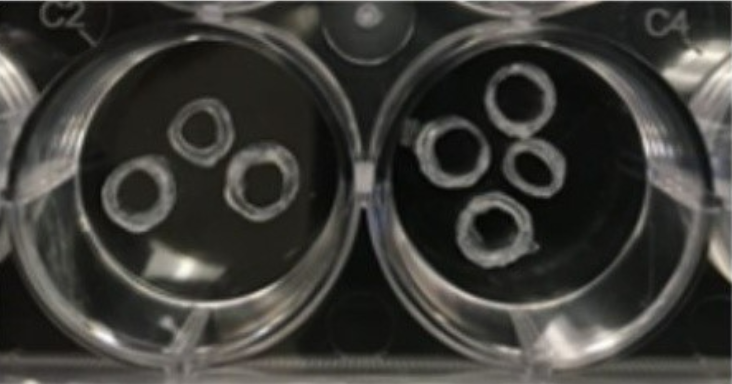
To create skeletal structures that are porous and stimulate cell growth, the researchers developed reusable circular templates and molds. Hydrogel skeletal structures were flexible and saturated with various concentrations of strontium, which was released initially within 24 hours and then sustained for four days with minimal toxicity.
Laboratory tests conducted showed that strontium-saturated skeletal structures increased the activity of isolated gingival fibroblast cells, while the hydrogel skeletal structure had only a minor impact on these cells. The full report on the results and research methods can be found in the article titled – “Strontium‐loaded hydrogel scaffolds to promote gingival fibroblast function” published in the Journal of Biomedical Materials Research Part A, 2022; 111 (1): 6 DOI.
Other contributors to the research include University at Buffalo alumni Shahad Bakheet Al-Sharif, co-author and faculty member at King Abdulaziz University in Saudi Arabia; Rofida Wali, co-author and faculty member at Umm Al-Qura University in Saudi Arabia; and Bhoomika Sheth, a quantum dot manufacturing engineer at STMicroelectronics. The University at Buffalo research team also includes Mark Swihart, PhD, Professor and Chair of the Department of Chemical and Biological Engineering, and Kayven Chen, a chemical and biological engineering student, both from the School of Engineering and Applied Sciences; as well as Sebastiano Andreana, DDS, MS, Professor of Restorative Dentistry and Director of Implant Dentistry, Rosemary Dziak, PhD, Professor of Oral Biology, and Stephen VanO, Research Technician, all from the School of Dentistry at the University at Buffalo.


