Full-Arch Screw-Retained Implant-Supported Restoration on a Bar with All-on-4 Concept, Accompanied by Tooth Extraction in a 65-Year-Old Male Patient
Abstract:
At the time of treatment, the patient had severely impaired masticatory function and a very poor aesthetic condition of his smile. Most of the teeth were missing, the 14 that remained could not be restored. In addition, gingival recession and severe periodontitis were observed. It was decided to extract the remaining teeth and place four implants in each jaw. The restoration was to be carried out according to the delayed protocol. During the period of osseointegration, the patient used removable dentures. CAD/CAM technologies were used in manufacturing of the permanent prosthesis. The prosthesis itself was acrylic and bar-supported. As a result, the masticatory function was completely restored and excellent aesthetics was achieved.
The value of this case for the dental community
Despite the clinical complexity of this case, a good result was achieved, so this case may be insightful for specialists interested in full-arch restoration, including using the all-on-4 and all-on-6 protocols, as well as for those interested in the features of screw retention of prostheses on multi-unit abutments.
Patient History
The patient is a 65-year-old man. Upon examination, loss of numerous teeth was discovered. The remaining teeth were scattered and could not be restored, and the clinical picture was complicated by periodontitis. The patient complained of pain and significant discomfort, as well as the inability to eat properly.

Clinical Examination
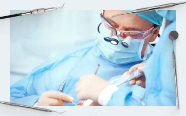
During the examination and throughout the course of treatment, a photo protocol was followed; the photos below show the initial condition.
")
Initial clinical picture of the patient (photo protocol)
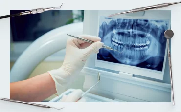
The orthopantomogram shows the condition of the bone tissue, and it is clear that there is a sufficient volume of bone tissue for placement of implants only in the anterior sections of the upper and lower jaw.
 Of Both Jaws")
X-ray (orthopantomogram) of both jaws
Diagnosis
Significant carious damage to the remaining teeth, the situation is complicated by periodontitis
Teeth are subject to removal with subsequent restoration of the dental arch with support on implants.
Treatment Plan
Based on the examination and photographs, it was decided to restore both dental arches using 4 implants. This option was considered optimal because:
- It restores masticatory function and quality of life by 100% compared to removable vacuum-formed protheses.
- It does not require surgery to increase the volume of bone tissue, which would be needed to place six or more implants. Every surgery increases the risk of complications, so it is always better to manage with a minimum number of surgeries.
Considering the shape of the patient’s jaws, placement of all implants is planned with a distal inclination.
The screw-retained multi-unit abutments-supported type of prosthesis was chosen, based on:
- Recommendations presented at ITI Consensus Conference Bern 2013.
")
Here are the quotes from the overall findings and recommendations of Dr. Wittneben’s study:
“The total event rate for biologic complications was significantly higher with cemented compared to screw-retained reconstructions.
Presence of fistula/suppuration appeared statistically significantly more often with cemented reconstructions.”
“Considering the risks with cemented reconstructions and the limited options for interventions after definitive cementation, it seems to be appropriate to recommend a preference towards screw retention of implant-supported reconstructions.”
2. Consensus Statements and Recommended Clinical Procedures Regarding Restorative Materials and Techniques for Implant Dentistry Collected and published after the abovementioned consensus conference.
")
Screw retention may be recommended:
- In situations of minimal interarch space
- To avoid a cement margin and thus the possibility of cement residue This is especially important for multiple implantations, where it is extremely difficult to clean off remove cement residue, and contact of cement residue with soft tissues significantly increases the risk of inflammation for patients who have had episodes of peri-implantitis.
- When retrievability is of importance
- In the esthetic zone, to facilitate tissue contouring and conditioning in the transition zone (emergence profile)
- To facilitate screw retention, it is recommended that the implant be placed in a prosthetically driven position
Since it is not possible to place the implants in an ideal position due to the anatomical features of the patient’s jaws, it was decided to compensate for the non-optimal angle by installing angled abutments.
It is planned to use CAD/CAD systems to design and manufacture the restoration, which will speed up the process and reduce the number of fittings and adjustments of the restoration.
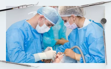
Treatment Implementation
After diagnostics and treatment plan development, treatment began. At the first stage, the following teeth were removed:
11; 12; 13; 15; 17; 22; 23; 27; 31; 32; 33; 42; 43; 44.
Simultaneously with the extraction of teeth, the patient was fitted with Uniqa Dental implants.

Implant placement procedure
The following types of implants were selected and installed:


At the next stage, haling caps were installed into the implants, and the soft tissues were sutured. A removable prosthesis was fabricated for a four-months osseointegration period.
After healing and gingival cuffs formation, scan abutments were installed in the implants and the intraoral space was recorded with an intraoral scanner.

Installation of scan bodies at the implant level
Based on the data obtained, it is possible to create a 3D model of the jaws on which the position of the implants and the exit angles of the screw shaft will be accurately marked. The specialist uses this information to determine the type of multi-unit abutments. Important parameters are gingival height and MUA angle. Uniqa Dental has two abutment angle options: 17° and 30°, each modification is available in three gingival heights: 1; 2; 3 mm.


After processing the virtual model and selecting the right abutments from the digital library, the selected multi-unit abutments were installed in the implants, see photo.


The next stage is installation of scan bodies at the abutment level and a new recording with an intraoral scanner to obtain information about the exit angles of the abutment screw shafts. Based on these data, a prosthesis with an anatomically correct line of occlusion will be designed. If possible, you should avoid situations where the screw shaft extends onto the buccal side of the restoration, especially in the esthetic zone, but sometimes this is unavoidable and you need to ensure that the screw shaft is at least covered with your lips when smiling.

Installed scan bodies at the abutment level for recording with an intraoral scanner
Based on the obtained 3D model of the jaws, specialists develop a prosthesis; in this case, a cobalt-chromium alloy was used as a frame, on which an acrylic restoration was formed.
The prosthesis was successfully manufactured and placed in the patient’s oral cavity.

Restoration aesthetics

Control X-ray 18 months later
Summary table of the clinical case
| Parameter | Result |
| Implant status | Osseointegration is complete |
| Healing phase | Full recovery |
| Bone tissue status | No bone loss |
| Stability | Good implant stability |
| Aesthetics | Good |
| Restoration status | Screw retention – several full arch units |
References
ITI Consensus Conference Bern 2013

-
Hot
10 Х 17° and 30° Angled Multi-Unit Abutment D-Type Internal Hex Regular Platform
Original price was: $680.$578Current price is: $578. Buy Now -
Hot
10 Х Angled Multi-Unit Abutment D-Type for Megagen AnyRidge® C
Original price was: $680.$578Current price is: $578. Request -
Hot
10 Х Angled Multi-Unit Abutment D-Type for MIS® V3 C
Original price was: $680.$578Current price is: $578. Buy Now -
Hot
10 Х Angled Multi-Unit Abutment D-Type for NobelActive® C
Original price was: $680.$578Current price is: $578. Buy Now